On-line Guide to Diagnostic Electron Microscopy

The goal of this first-aid manual, designed mainly for use by The Fingerland Department of Pathology, was to offer students and starting electron microscopists specializing in diagnostics of human diseases a quick visual overview of the ultrastructural appearance of some basic pathological changes. It is far from being extensive enough to embrace the whole range of this examination method. Users of our atlas are assumed to already be acquainted with basic ultrastructural cytology and histology of organ systems. This guide is not a textbook. The figure captions are minimalized and will be updated, completed or corrected in time. For further extensive analysis of structures we urge you to consult, for example,
Jan Vincents Johannessen:
Electron microscopy in human medicine series
McGraw-Hill, 1978-1985,
Ann M. Dvorak, Rita A. Monahan-Earley:
Diagnostic Ultrastructural Pathology. Vol. I - III,
CRC Press, 1995,
Feroze N. Ghadially:
Ultrastructural Pathology of the Cell and Matrix
Fourth edition, Vol. I and II., Boston, Butterworth–Heinemann, 1997,
G. R. Dickerson: Diagnostic Electron Microscopy,
Springer, 2000
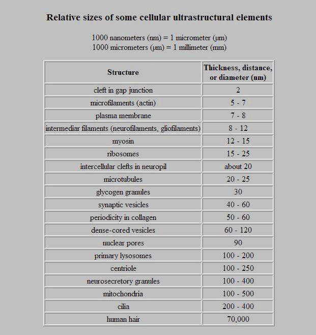
The content of this Guide is a collection of some cases only, examinated during several decades in our laboratory. About 1 500 of electron micrographs, on purpose often repeated in many variations, may serve to show a variability in details and developmental stages of a disease. Histopathologists and even more the electron microscopists, must take into consideration during their decisions also artifacts caused by an inadequate fixation and a mechanical damage during a withdrawal of a tissue and an autolysis in samples from autopsies. Some of these are shown in our figures, too. Histopathologists have magnification scales firmly imprinted into their memories and therefore their published pictures are usually free of scales. A short table of sizes of some ultrastructural elements is attached.
This Guide was edited by Josef Spacek and in its preparation took part Ladislav Kubes, Milan Resl, Mirek Podhola and Eva Hovorkova. The authors thank their clinical colleagues for a supplementation with bioptic and autoptic samples and laboratory technicians for the processing.
The samples of which micrographs are presented in this Guide were withdrawn from experimental animals for reasons of research in accordance with the University Medical Ethics Committee or from human tissues withdrawn with informed consent for diagnostic or therapeutical reasons.
The presented figures are not containing any private data and the authors have no objections against their non-commercial use. In such a case, the source should be refered as follows:

Josef Spacek, http://www.patologie.info/ogdem/
Address for correspondence:
Prof. MUDr. Josef Spacek, DrSc, FRMS
Fingerland Department of Pathology
Charles University Hospital
500 02 Hradec Kralove
Czechia
spacekjos702@gmail.com


