Úvod On-line Guide to Diagnostic Electron Microscopy 4. Oral cavity, salivary glands
On-line Guide to Diagnostic Electron Microscopy

F,23y. | normal oral mucosal epithelium

F,23y. | normal oral mucosal epithelium - hemidesmosomes and anchoring fibrils
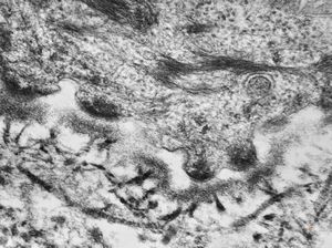
F,23y. | normal oral mucosal epithelium - hemidesmosomes and anchoring fibrils
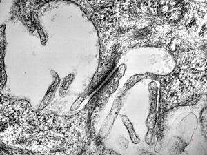
F,23y. | normal oral mucosal epithelium - desmosom
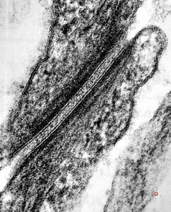
F,23y. | normal oral mucosal epithelium - desmosom
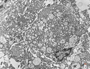
normal seromucinous salivary gland
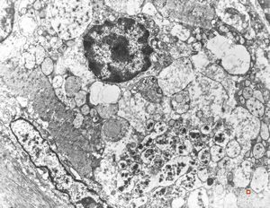
normal seromucinous salivary gland
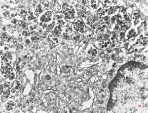
normal seromucinous salivary gland
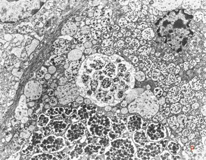
normal seromucinous salivary gland
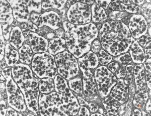
normal seromucinous salivary gland
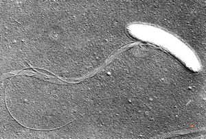
oral saprophytic bacterium
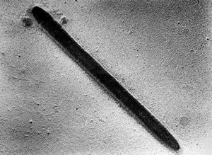
oral saprophytic bacterium
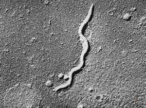
oral saprophytic bacterium
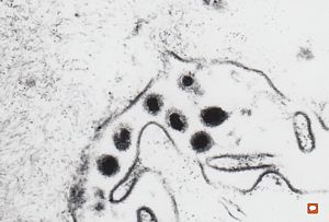
F,65y. | Epstein-Barr or cytomegaly virus - hairy leukoplakia - tongue
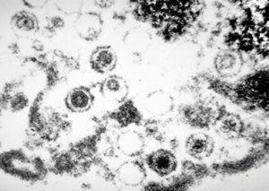
F,65y. | Epstein-Barr or cytomegaly virus - hairy leukoplakia - tongue
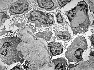
M,46y. | nuclear pseudoinclusion in plasmocyte - susp. amyloid … tonsillar tumor … Waldenström macroglobulinemia
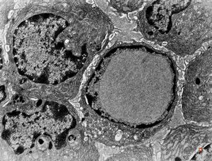
nuclear pseudoinclusion in plasmocyte
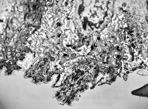
M,7y. | epidermolysis bullosa (dystrophic type) - oral mucosa
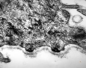
M,7y. | epidermolysis bullosa (dystrophic type) - oral mucosa
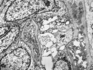
M,52y. | cylindroma - salivary gland
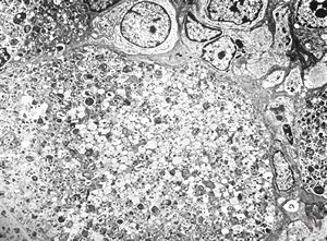
F,1y. | epulis congenitalis (granulomatosa)
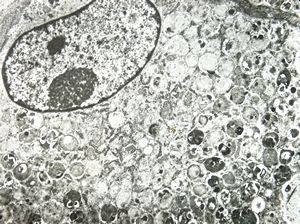
F,1y. | epulis congenitalis (granulomatosa)
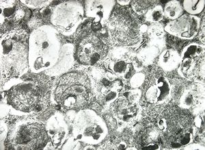
F,1y. | epulis congenitalis (granulomatosa)
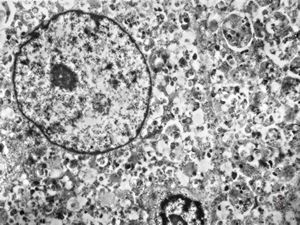
F,1y. | epulis congenitalis (granulomatosa)
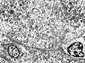
F,1y. | epulis congenitalis (granulomatosa)
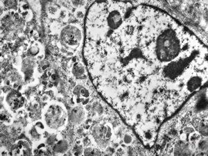
F,1y. | epulis congenitalis (granulomatosa)
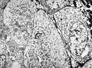
M,58y. | carcinoma gl. parotis
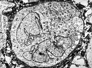
M,58y. | carcinoma gl. parotis
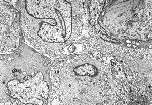
F,22y. | Pindborg tumor
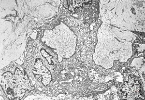
F,22y. | Pindborg tumor
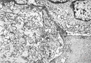
F,22y. | Pindborg tumor
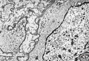
F,22y. | Pindborg tumor
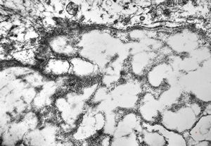
F,22y. | Pindborg tumor
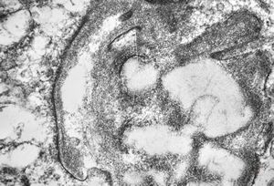
F,22y. | Pindborg tumor
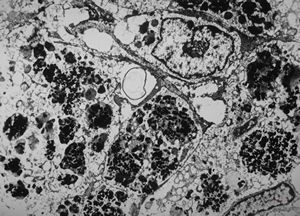
M,30y. | melanom - gl. parotis
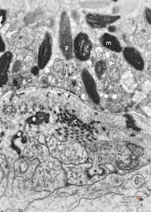
M,6m. | maxilla - melanotic progonoma (melanotic neuroectodermal tumor of infancy)
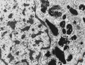
M,6m. | maxilla - melanotic progonoma (melanotic neuroectodermal tumor of infancy)
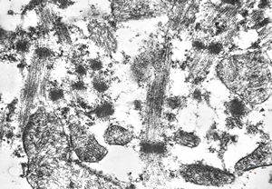
F,75y. | rhabdomyoma - tongue
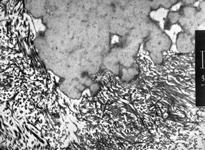
F,74y. | ossifying fibromyxoid tumor - submandibular gland
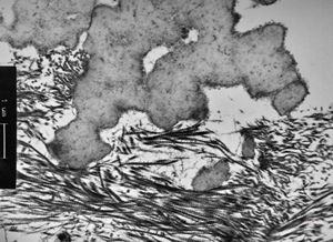
F,74y. | ossifying fibromyxoid tumor - submandibular gland
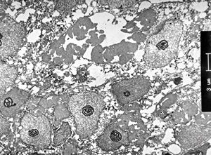
M,61y. | carcinoma - submandibular gland
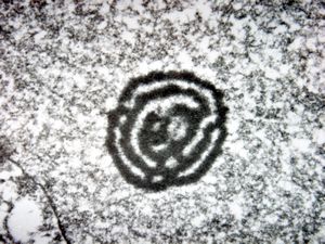
M,61y. | carcinoma - submandibular gland - activated nucleolus
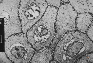
M,66y. | follicular cyst with hyaline Rushton bodies
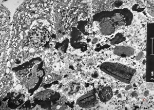
M,66y. | follicular cyst with hyaline Rushton bodies
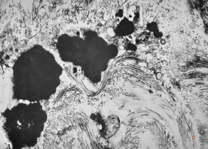
M,66y. | follicular cyst with hyaline Rushton bodies
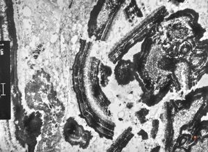
M,66y. | follicular cyst with hyaline Rushton bodies
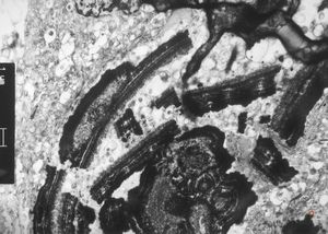
M,66y. | follicular cyst with hyaline Rushton bodies
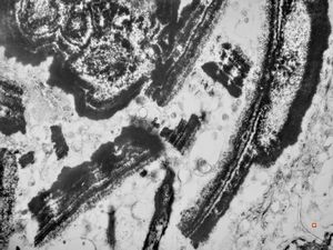
M,66y. | follicular cyst with hyaline Rushton bodies
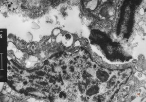
M,66y. | follicular cyst with hyaline Rushton bodies
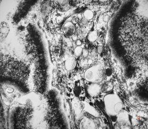
M,66y. | follicular cyst with hyaline Rushton bodies
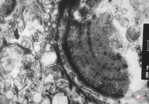
M,66y. | follicular cyst with hyaline Rushton bodies
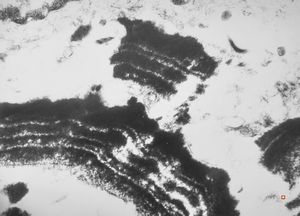
M,66y. | follicular cyst with hyaline Rushton bodies
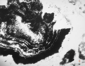
M,66y. | follicular cyst with hyaline Rushton bodies
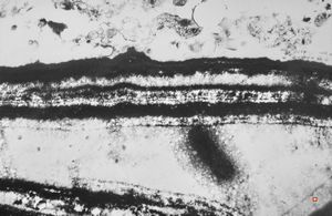
M,66y. | follicular cyst with hyaline Rushton bodies
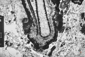
M,66y. | follicular cyst with hyaline Rushton bodies
Download PDF with higher resolution
Contents
- Introduction
- 1. Cell and matrix pathology, unusual structures, artifacts
- 2. Infectious agents (viruses, bacteria, fungi, parasites)
- 3. Respiratory tract
- 4. Oral cavity, salivary glands
- 5. Gastrointestinal tract
- 6. Liver, gallbladder, pancreas
- 7. Cardiovascular system
- 8. Blood, bone marrow, spleen, lymphatic system
- 9. Urinary tract
- 10. Male reproductive system
- 11. Female reproductive system
- 12. Breast
- 13. Skin
- 14. Bone and joints
- 15. Soft tissues (connective tissue, muscles, peripheral nerves)
- 16. Endocrine system
- 17. Inborn metabolic disorders
- 18. Eye and ear
- 19. Central and peripheral nervous system
- 20. Atlas of Ultrastructural Neurocytology


